Friday 26 February 2016

PERFORMING A TWO-MINUTE HANDWASH


a. Purposes.

(1) Prevent nosocomial infection.

(2) Maintain safe, clean environment for patients.

(3) Provide safety for health care workers.

(4) Prevent cross-contamination of patients or the spread of microorganisms.


b. Scope of Responsibility.
Teaching patients and visitors about procedures and appropriate times for handwashing is an important role for the health care provider. This enables the patient and family to inhibit the spread of microorganisms when health care is continued at home.

The importance of handwashing before and after handling food, after handling contaminated articles, and before and after elimination should be stressed in the teaching process.

c. The Two-Minute Handwash.

A two-minute handwash will provide appropriate protection before you begin working with a patient. A 30-second handwash should be sufficient before caring for another patient. A one-minute handwash should be appropriate if you have handled organic material or a contaminated object.

d. Additional Actions.

In addition to handwashing, other actions can be taken to reduce the chance of transmitting microorganisms. The patient should receive a personal set of care articles, such as a bedpan, urinal, bath basin, thermometer, water pitcher, and drinking glass to prevent cross-contamination. Articles such as contaminated equipment and soiled linen should be placed in special waste containers or laundry bag, and kept away from your uniform.

e. Schedule for Handwashing.

Handwashing is essential:

(1) Before and after caring for a patient.

(2) After contact with organic material, such as feces, wound drainage, and mucous.

(3) In preparation for an invasive procedure such as suctioning, catheterization, or injections.

(4) Before performing a dressing change or contact with an open wound.

(5) Before preparing and administering medications.

(6) After removing disposable gloves or handling contaminated equipment.


STEPS FOR PERFORMING THE TWO-MINUTE HANDWASHING


a. Step One.
(1) Inspect hands, observing for visible dirt, breaks, or cuts in the skin and cuticles (figure 5-1).
(2) Determine contamination of hands.
(3) Assess areas around the sink that are contaminated or clean.
(4) Explain to the patient the importance of handwashing.

Figure 5-1. Inspecting hand.

b. Step Two.
(1) Remove all jewelry (except plain wedding band) and push watch and long sleeves above wrists.
(2) Adjust water to right temperature and force (figure 5-2).
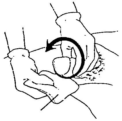
Figure 5-2. Adjust water temperature and force.

c. Step Three.
(1) Wet hands and wrists under the running water, always keeping hands lower than the elbows.
(2) Lather hands with liquid soap (about one teaspoon).
(3) Wash hands thoroughly using a firm circular motion and friction on back of hands, palms, and wrists. Wash each finger individually, paying special attention to areas between fingers and knuckles by interlacing fingers and thumbs, and moving fingers back and forth.
(4) Wash one minute, rinse thoroughly, relather, and wash another minute, using a continuous amount of friction.
(5) Rinse wrists and hands completely, keeping hands lower than elbows (figure 5-3).

Figure 5-3. Hands lower than elbow.

d. Step Four.
(1) Clean the fingernails carefully under running water, using fingernails of other hand or blunt end of an orange stick.
(2) Dry hands thoroughly with paper towels. Start by patting the fingertips, hands, and then wrists, and forearms.
(3) Turn off faucets with a dry paper towel (figure 5-4).
(4) Use hand lotion if desired.
(5) Inspect hands and nails for cleanliness.

Figure 5-4. Turning off water with towel.

COLLECTING SPUTUM


a. General.

Sputum is mucus from the lung.

A sputum specimen must come from deep in the bronchial tree. Expectoration from throat and mouth secretions cannot be used as a sputum specimen. Early morning is the best time to collect a sputum specimen because the patient has not yet cleared the respiratory passages.


Many tests can be performed on sputum, such as a culture and sensitivity, cytological examination, and test for acid-fast bacillus. Some patients cannot expectorate a specimen and must have a pharyngeal suctioning to obtain sputum. Closed-method collection containers protect you from contamination from body fluids. The medical specialist explains the procedure and prepares the patient for the test.

b. Important Points.

(1) Oral hygiene should be provided after the procedure for patient comfort.

(2) Accuracy of test decreases if delivery of specimen to laboratory is delayed.

(3) Make certain the patient knows how to perform sputum collection.

(4) The nurse must be prepared to obtain the specimen by suctioning if the patient cannot cough.

c. Procedure.

(1) Read physician’s orders.

(2) Collect supplies.

(3) Introduce yourself.

(4) Identify the patient by identification band.

(5) Explain procedure to patient.

(6) Wash hands and don gloves.

(7) Position patient in Fowler’s position.

(8) Instruct patient to take three breaths and force cough into sterile container.

(9) Attach laboratory requisition.

DETERMINING PRESENCE OF OCCULT BLOOD IN STOOL


a. General.
The presence of blood in body waste is abnormal.
Blood in the stool may be bright red, which indicates that the blood is fresh and that the site of bleeding is in the lower gastrointestinal tract. On the other hand, black-tarry-feces means the presence of old blood and that the site of bleeding is higher in the gastrointestinal tract.


When blood is present in the stool but cannot be seen without the use of a microscope, it is referred to as occult or hidden. A hemoccult test detects occult blood in feces.
b. Important Points.
(1) Do not confuse hemorrhoidal bleeding with upper gastrointestinal bleeding.
(2) Meat-free diet may be ordered 3 days before the test.

COLLECTING A TWENTY-FOUR HOUR URINE SPECIMEN


a. General.
Some tests require that the entire volume of urine from a 24-hour period be collected. The procedure for ensuring that the test can be performed accurately should be followed carefully.


b. Important Points.
Use strict sterile technique to prevent infection in the urinary system. Insert the catheter gently to prevent pain or discomfort, as catheterization should not be painful. Teach the patient to relax by deep breathing during catheterization. Answer the patient’s questions about the procedure.
c. Procedure.
(1) Read physician’s order.
(2) Wash hands.
(3) Identify the patient.
(4) Post “Do not disturb” signs on patient’s door, bathroom door, and near patient’s bed.
(5) Explain procedure.
(6) Instruct patient about the importance of collecting all urine for 24 hours.
(7) Instruct patient not to place toilet tissue or fecal material in urine.
(8) Have patient void when the 24-hour specimen collection is to begin; discard this voiding.
(9) Place labeled container on ice if required. (Some agencies require refrigeration of all specimens. Others advocate that the urine container be placed on ice. For some collection procedures, such as the creatinine clearance test, refrigeration may not be necessary.)
(10) Save all urine for the 24-hours, then place each voided specimen into the larger container with preservative.
(11) Instruct patient to void a few minutes before the end of 24 hours; this urine is part of the 24-hour specimen.
(12) Send specimen to lab promptly; be certain label includes date and time specimen started, patient’s name, room number, and test ordered. If more than one container is necessary, make certain both are labeled and numbered.

COLLECTING A MIDSTREAM URINE SPECIMEN


a. General.

A midstream specimen is a voided specimen collected under conditions of thorough cleanliness after approximately the first 30 ml of urine has been voided.

The advantage of collecting a voided specimen in this manner is that if organisms appear in the urine, they are mostly from structures such as the bladder or kidneys rather than just surface contamination. Cleansing removes organisms from the urinary meatus. Voiding moves any residual organisms present in the urethra out with the beginning stream of urine.


b. Important Points.

Specimens of urine should not be allowed to stand at room temperature before they are sent to the laboratory. Bacterial growth is likely to occur as well as alter other results of the urinalysis.

The usual procedure is to store an aurum (gold) specimen in a refrigerator, if it is not taken directly to the laboratory. Specimens that are collected from multiple voidings are either refrigerated on the nursing unit or placed in a container with a chemical preservative.

c. Procedure.

(1) Read physician’s orders.

(2) Collect supplies.

(3) Introduce yourself to the patient.

(4) Identify patient by identification band.

(5) Explain procedure to patient.

(6) Wash hands and don clean gloves.

(7) If patient is able, allow patient to cleanse perineum with antiseptic solution. Separate the labia well on a female patient. Retract foreskin of an uncircumcised male. Use each cotton ball that is saturated with antiseptic solution one time only. If patient is unable to cleanse area, the nurse will assist with procedure.

(8) Assist the patient.

(a) Begin to void into container about 30 ml; then place the sterile specimen container so the sides of the labia of the female do not touch;

(b) To stop flow, void a small amount into specimen cup; and

(c) Without stopping flow, finish voiding into toilet seat collector.

(9) Secure the lid on the container.

(10) Cleanse and return toilet seat collector, if applicable.

(11) Label specimen appropriately.

(12) Ensure that specimen is taken to laboratory with requisition.

COLLECTING A STERILE URINE SPECIMEN


a. General.

A sterile urine specimen can be obtained either by inserting a straight catheter into the urinary bladder and removing urine or by obtaining a specimen from the port of an indwelling catheter using sterile technique.

Urine from the dependent drainage bag should not be used for a specimen, since it is not fresh and would not reflect accurate test results.


Residual urine, urine left in the bladder after voiding, can be measured at the time of catheterization. The patient voids, and catheterization is performed within 10 minutes. If more than 60 ml of urine remains in the bladder, this is residual urine and the patient may need to have an indwelling catheter inserted.
The medical nurse must prepare the patient by explaining which type of urine specimen will be collected. It is important to relieve any anxiety by assuring the patient that there should be no discomfort during the procedure if the patient will remain relaxed: the patient should experience only mild pressure as the catheter is inserted and will feel nothing when urine is collected from the catheter port.
b. Important Points.
(1) Have all supplies ready for the patient to perform the procedure.
(2) Make certain the patient understands the proper procedure for collecting the urine specimen.
(3) Be certain the specimen is labeled correctly: patient’s name, room number, date, physician, and type of specimen.
c. Procedure.
(1) Read physician’s orders.
(2) Collect supplies.
(a) Sterile cotton balls.
(b) Antiseptic.
(c) Sterile specimen container.
(3) Introduce yourself to the patient.
(4) Identify patient by checking his identification band.
(5) Explain the procedure to the patient.
(6) Obtain the catheter port collection:
(a) Clamp tubing just below catheter port for about 30 minutes (figure 4-1).
(b) Return in 30 minutes and clean the port with alcohol prep.
(c) Insert needle into port at 30-degree angle, and withdraw 5 to 10 ml of urine for a specimen (figure 4-2).

Figure 4-1. Clamp catheter port.
Figure 4-2. Insert needle into catheter port.

(d) Place urine in sterile specimen cup.
(e) Unclasp catheter.
(f) Label specimen, and send to laboratory with requisition.
(g) Document the procedure.
(7) Obtain straight catheter collection.
(a) Wash your hands and don sterile gloves, and prepare supplies, using sterile technique–wrap the edges of the sterile drape around the gloved hands.
(b) Place sterile drape under patient’s buttocks (figure 4-3).

Figure 4-3. Place sterile drape under buttocks.

(c) Open the lubricant container; add antiseptic (usually iodine solution) to the cotton balls.
(d) Lubricate the catheter about 1.5 to 2 inches (3.5 to 5 cm).
d. Catheterize the Female Patient.
(1) To expose the meatus, place the thumb and forefinger of the nondominant hand between the labia minora. Spread and separate upward. Consider the gloved hand that has touched the patient to be contaminated (figure 4-4).

Figure 4-4. Expose the meatus.

(2) Maintain the position of the contaminated hand until urine is flowing.
(3) Pick up the forceps and secure a cotton ball saturated with antiseptic solution–use one cotton ball for each stroke.
(4) Bring the cotton ball down the center over the meatus towards the rectum; next cleanse each lateral area from superior to inferior.
(5) Deposit used cotton balls onto plastic cover.
(6) To insert a catheter into a female with sterile gloves pick up catheter and insert through urinary meatus 2 to 3 inches (5 to 7.5 cm). DO NOT FORCE ENTRY OF THE CATHETER. Discontinue the treatment if the patient has unusual discomfort or if there is continual resistance to the insertion of the catheter. Report the information promptly.
(7) When urine flows, place end of catheter in specimen cup.
(8) Place lid on urine cup and label; clean up supplies.
(9) Send specimen to lab with requisition and document the procedure.
e. Catheterize the Male Patient.
(1) To cleanse the penis, swab the center of the meatus outward in a circular manner. Continue, using a new cotton ball for each progressively larger circle (figure 4-5).

Figure 4-5. Cleanse the penis.

(2) To insert a catheter into a male, apply gentle traction and pull the penis straight up; slightly pinch the end of the penis and insert the catheter 15 to 20 cm (6 to 8 inches). To facilitate the more difficult passage through the male urethra, ask the patient to breathe deeply; then rotate the catheter slightly. DO NOT FORCE ENTRY OF THE CATHETER. Discontinue the treatment if the patient has unusual discomfort or if there is continual resistance to the insertion of the catheter. Report the information promptly.
(3) When urine flows, place end of catheter in specimen cup.
(4) Place lid on urine cup and label. Clean up supplies, send specimen to lab with requisition, and document the procedure.

MAKING THE PATIENT OCCUPIED BED


a. General.
Changing bed linen and making a comfortable, neat bed while it is occupied by a patient usually follows the completion of a cleansing bath.
During this time, excellent opportunities are provided to establish good relations with the patient through patient-centered conversation and for instructing the patient how to move, turn, conserve energy, and maintain good body alignment.


If the patient is helpless or unconscious, two individuals should work together. The operator gives instruction and performs the procedure while the assistant holds the patient and helps to turn him. When an assistant is unavailable to assist a helpless patient, the side rails of the bed opposite the operator should be raised to prevent the patient from falling out of bed.
b. Precautions in Making a Patient Occupied Bed.
Some precautions in making a patient occupied bed are to prevent exposing the patient, provide for his safety, and (by the proper handling of linen) prevent the possible spread of microorganisms.
c. Equipment.
The following equipment should be obtained as required.
(1) Washbasin containing an appropriate solution.
(2) Cleaning cloth.
(3) Two sheets.
(4) One pillowcase.
(5) Protective sheet and cotton drawsheet as necessary.
(6) Paper bag.
(7) Clothes hamper.
d. Procedure.
The following procedure for making a patient’s occupied bed is appropriate when the patient is not helpless.
(1) Step 1.
(a) Remove the pillow and use the crank handle to level the bed if permitted.
(b) Loosen the bed linen while moving around the bed, slightly raise the mattress and lift the linen edges free. To prevent the linen from snagging on the springs, do not tug or jerk it.
(c) Pull mattress up to the head of the bed as necessary.
(d) Remove the spread and blanket, leaving the top sheet as a cover for the patient.
(e) Assist the patient to turn toward you, to the side of the bed, keeping his body covered with the sheet. If required, raise and latch the bedrail when the patient’s position has been adjusted.
(2) Step 2.
(a) Go to the opposite side of the bed. Place the chair in a convenient location.
(b) Roll all bedding in layers close to the patient’s back. Smooth and tighten the mattress cover from top to bottom.
(c) Place the clean foundation (bottom) sheet on the exposed section of the mattress with the hem seam down and the centerfold in the midline of the bed. It should be folded against the patient, bottom edge even with the foot of the mattress. Tuck under the top edge, miter the corner, and smooth and tuck the side under, moving from head to foot.
(d) Replace the protective sheet, if used. Hold the linen folds in place in the center of the bed with one hand, and bring the rolled protective sheet back over the linen folds to the clean foundation. Place the clean cotton drawsheet over the protective sheet, rolling the excess folds toward the patient.
Smooth and tuck under the sides of both sheets to complete the near side of the foundation. (The cotton drawsheet should completely cover the protective sheet in order to prevent irritation of the patient’s skin, when it is exposed to either the rubber or laminated cotton sheet.)
(e) Tuck all linen folds under the patient as smoothly as possible. Assist the patient to roll over the linen folds to the clean foundation side.
(3) Step 3.
(a) Secure the side of the bed, if needed, before going to the opposite side of the bed.
(b) Go to the opposite side of the bed. Pull the rolled linen through. Keep the clean linen close to the patient’s back; remove the soiled linen and place it in the clothes hamper. Tighten the mattress cover, head to foot. Complete the foundation.
NOTE: If protective sheets (or drawsheet only) are used, pull taut and wrinkle free by tightening the center portion first, then the upper and lower ends.
(c) Turn the patient to the center of the bed. Center the clean top sheet over the patient. Instruct the patient to hold the clean top sheet while you remove the soiled top sheet from underneath, pulling gently from top to bottom. Place the soiled sheet in the clothes hamper.
(d) Replace the blanket and spread, instructing the patient to check for free movement of his feet to be sure the top bedding is loose enough before tucking under and maturing the corners at the foot.
(e) Complete the cuff at the head of the bed. Fold down the top bedding to a level comfortable for the patient.
(4) Step 4.
(a) Place a clean case on the pillow. Replace the pillow under the patient’s head. Use the crank handle at the foot of the bed and adjust as needed. Also adjust the sides of the bed as needed.
(b) Place the bedside cabinet and the signal cord within the patient’s reach.
(5) Step 5.
(a) Damp-dust the unit. Attach the clean paper bag and place towels and washcloth in the proper place.
(b) Remove all unnecessary equipment and articles from the unit. Leave the unit clean and orderly.
(c) Return the clothes hamper to the storage area. Discard the waste. Wash and sanitize the equipment that is returned to the utility room. Wash your hands.